Fetal pleural effusion is an accumulation of fluid in the chest cavity of a developing fetus. Fetal pleural effusion is an abnormality resulting from accumulation of fluid in the chest cavity and the condition was first described by Carroll in 1977 1.

Fetal Echocardiography A Large Pericardial Effusion White Star Download Scientific Diagram
Hydrops fetalis is severe swelling edema in an unborn baby or a newborn baby.
. He was ventilated from birth for 16 days. The heart is surrounded by a double-layered sac-like structure called the pericardium. The pericardial sac is composed of the thin visceral pericardium which consists of a single layer of cells adherent to the cardiac epicardium and the thicker fibrous parietal pericardium composed of collagen and elastin which is adherent to the lungs.
Pericardial effusion requires attention because of its underlying pathology and its potential for producing hemodynamic compromise. Pericardial effusion is often related to inflammation of the pericardium due to disease or injury but pericardial effusion can. Hydrops fetalis HIGH-drops fee-TAH-lis is a life-threatening condition in which abnormal amounts of fluid accumulate in two or more body areas of an unborn baby.
Fetal pleural effusion is a rare condition with a reported incidence ranging from 110000 to 115000 2 4. As the fluid increases it can compress the developing lungs and heart. Post-cardiotomy syndrome tends to occur within 1-2 weeks of the surgery.
Idiopathic pericarditis accounts for 37 to 68 of inpatient admissions for pericardial effusions or acute pericarditis in children. Pericardial effusion in children. Many forms of pericardial diseases can occur sporadically in pregnant women.
A pericardial effusion is excess fluid between the heart and the sac surrounding the heart known as the pericardium. BabyCentre may earn a commission from shopping links. Hydrops fetalis is a condition of excess fluid accumulation in the fetus.
1 2 3 The usual approach to initial management of acute pericarditis includes administration of nonsteroidal antiinflammatory agents. Slightly elevated blood pressure andor non-specific ST-T changes in association with pericardial effusion. Fetal heart failure leads to pericardial effusion.
In pericardial effusions both lungs are compressed towards the back of the chest. After losing my son to HLHS with aortic stenosis and endocardial fibroelastosis Im currently expecting our rainbow baby. Genetic testing for GACI can be confirmed through an amniocentesis or chorionic villus sampling CVS.
Colchicine has emerged as an important adjunct in management of. We reviewed pediatric patients with moderate or large effusions diagnosed at Childrens Hospital Boston. Pericardial effusion is the accumulation of excess fluid around the heart.
The effusion may be only on one side or on both sides. In pleural effusions the lungs are compressed towards the midline by the fluid within the pleural space. We reviewed the clinical data on 44 consecutive patients seen between 1986 and 1993.
Pericardial effusion on ultrasound may also indicate an increased risk of Down syndrome in a fetus 1. Pericardial fluid 2 mm or greater in thickness may be associated with structural anomalies or hydrops but its clinical significance in the absence of these associated findings has not been evaluated. Abrams et al 15 16 The causes of hydrops fetalis can be determined antenatally in 5085 of the cases.
It was our hypothesis that the etiology of and danger from large pericardial effusions are different at different ages. Hydrops develops when too much fluid leaves the babys bloodstream and goes into the tissues. Although the fluid buildup may appear anywhere in the babys body it most often occurs in the abdomen around the heart or lungs or under the skin.
It is a life-threatening problem. Infants and young children tend to be fussy and have decreased feeding with tachycardia being an important physical sign. Our study population included 52 fetuses with.
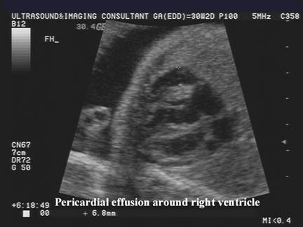
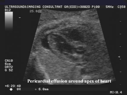
Excessive water and salt retention in subjects with a higher mean weight gain. Small pericardial effusion noted on the. In either case the size of the effusion can be monitored by measuring the distance between the chest.
The space between the layers normally contains a very small amount of fluid. Congenital heart disease and heart arrhythmias may cause fluid accumulation. We assessed the outcome in fetuses with isolated pericardial effusions of at least 2 mm thick.
Yadav DK Agarwal S Aier T Gupta V. A pericardial effusion refers to the accumulation of fluid in the pericardial sac surrounding the heart. It has several characteristic antenatal ultrasonographic findings such as skin edema pleural effusion pericardial effusion ascites and polyhydramnios.
Cysts infection and an abnormal opening between the abdomen and chest cavity can also lead to pericardial effusion. This is similar to a routine ultrasound but provides a more in-depth evaluation. The second baby boy was born at 26 weeks gestation with a birth weight of 880 grams.
The incidence of fetal pleural effusion in. The most frequent are. The outlook for each individual case depends on the amount.
The sonographer was lovley and said she doesnt think it is going to be anything to serious to worry about. It is important to remember that almost 50 of patients with symptomatic pericardial effusion and neoplastic disease have a nonmalignant cause such as radiation-related idiopathic infectious including tuberculous and fungal and lymphatic obstruction. The underlying cause of pleural effusion in a fetus may include genetic issues infection and heart or lung conditions.
Effusion size was determined in offline review of echocardiograms. My unborn baby has pericardial effusion. The objective of this study was to determine the contemporary etiologies treatment and outcomes of moderate and large pericardial effusions in pediatric patients.
Post mortem examination showed massive 18 ml pericardial effusion consisting of creamy fluid suggestive of parentral nutrition. The clinical presentation of pericarditis in children is similar to that of adults with certain caveats. Pericardial effusion fluid around the unborn babys heart If additional imaging is needed a level 2 ultrasound may be ordered.
There was no evidence of macroscopic cardiac rupture or perforation. - UPDATE O we had our 20 wk scan earlier and have been told that our baby has a pericardial effusion which basically means a small amount of fluid in the heart. I am 28 5 and todays cardiac scan shows the baby to have pericardial effusion fluid around the heart I also had bad news yesterday at a growth scan that our baby is measuring small.
Ultrasound Of Pericardial Effusion

Fetal Pericardial Effusion Radiology Reference Article Radiopaedia Org

Fetal Echocardiography At 11 13 Weeks Pericardial Effusion Youtube
Pericardial Effusion Sujyotheartclinic

Pericardial Effusion A 29 Week 5 Day Gestation Fetus With Aortic Download Scientific Diagram
Ultrasound Of Pericardial Effusion
Get The Facts About Fetal Hydrops Hydrops Fetalis

Fetal Pericardial Effusion Radiology Reference Article Radiopaedia Org
0 comments
Post a Comment